Figure 4.
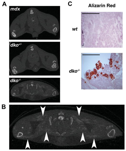
Ectopic calcification in the soft tissues of dko−/− mice. (A) 7-week-old mdx, dko+/−, and dko−/− mice were evaluated by micro-CT and representative images are shown to illustrate the presence of ectopic calcification (EC) in the proximal hind limb soft tissues of dko−/− mice exclusively. (B) A larger view of the micro-CT image from (A) with arrowheads demonstrating EC in a dko−/− mouse. (C) Skeletal muscle cryosections were prepared from the gluteus maximus of these mice and processed for histological analysis. Representative images obtained with alizarin red staining are shown. Scale bars in the upper left-hand corner represent 0.5 mm (at 4× magnification).

