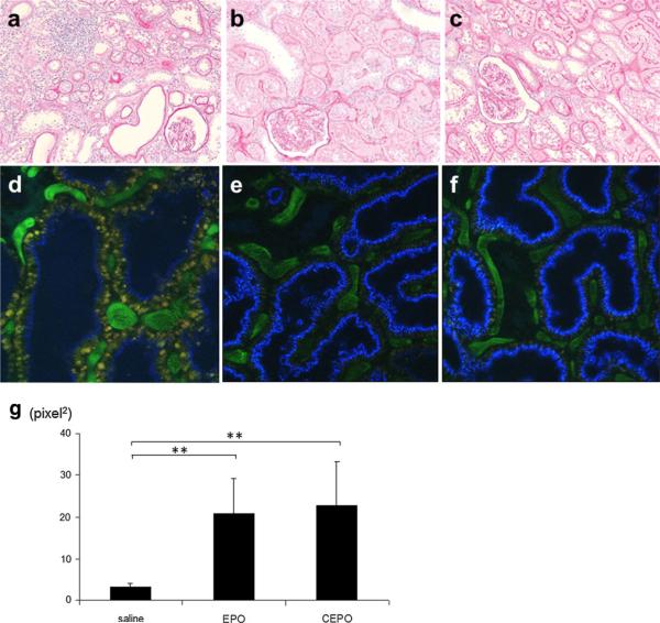
Fig. 2.
Representative morphological changes. PAS staining of the kidney sections obtained from remnant kidney treated with saline (a), EPO (b), or CEPO (c) for 8 weeks. Two-photon microscopy showed that saline-treated remnant kidney showed decreased endocytosis (blue) (d), while treatment with EPO (e) or CEPO (f) preserved endocytotic function (magnification ×400). The cascade blue-positive area was calculated as tubular endocytotic function (g) (**p < 0.01)

