
FIG. 5.
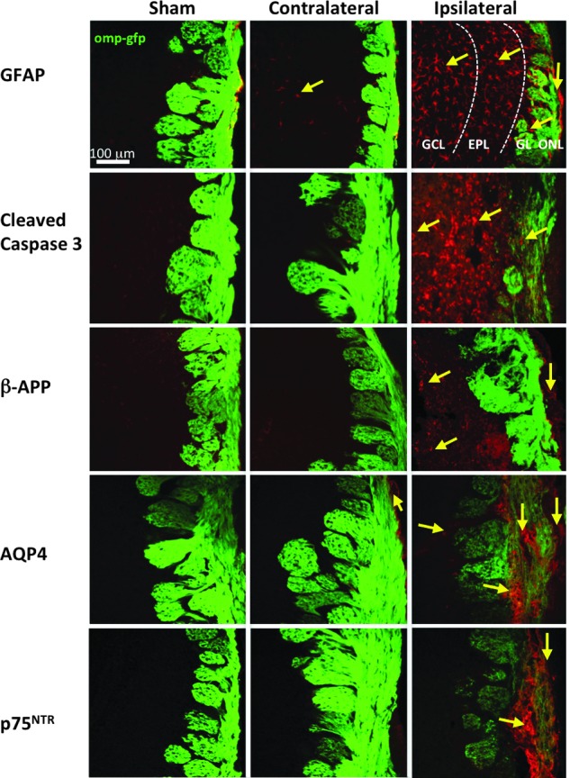
Markers of traumatic brain injury (TBI) in the olfactory bulb (OB) 4 days post-OBI (olfactory bulb impact). Representative images from sham, contralateral, and ipsilateral OB are shown from left to right. Glial fibrillary acidic protein (GFAP) (A), cleaved caspase-3 (B), beta amyloid precursor protein (β-APP) (C), aquaporin 4 (AQP4) (D), and p75NTR (E) showing a variable increases in TBI markers between ipsi- and contralateral OB with some changes extending into deeper layers. GCL, granule cell layer; EPL, external plexiform layer; GL, glomerular layer. Color image is available online at www.liebertpub.com/neu