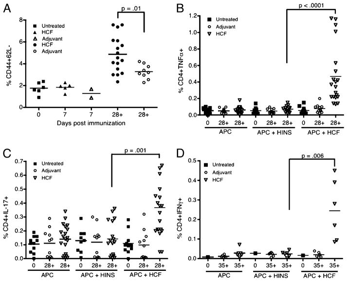
FIGURE 2.
HCF-immunized mice develop CD4 T cells with an activated effector phenotype (CD44high+CD62L−) and the ability to produce proinflammatory cytokines. DBA/1J mice, aged 6–8 wk, were untreated (Untreated), or immunized on days 0 and 21 with CFA (Adjuvant) or HCF in CFA (HCF), and draining LN were harvested at various time points (days 7, 28, or 35). (A) Surface staining was performed, followed by flow cytometric analysis, in which CD4+ cells were gated from live cells. The percentage of CD44high+ 62L− CD4 T cells is quantified for each group of mice with each shape representing one mouse. In (B)–(D), LN cell suspensions were stimulated with APCs alone as a negative control, APCs and irrelevant Ag (HINS), or APCs and HCF. After stimulation, cells were incubated with GolgiPlug for 4 h and then stained for intracellular cytokines. The percentage of (B) TNF-α+ CD4 T cells, (C) IL-17+ CD4 T cells, and (D) IFN-γ+ CD4 T cells is quantified for each group of mice. Relevant statistically significant differences are shown, and the bar represents the mean for each group at the respective time point.

