FIGURE 3.
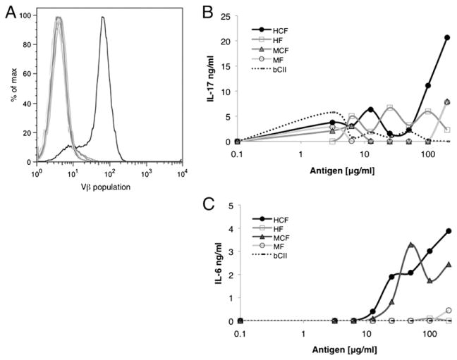
The HCF4Vβ7+ line is specific for CF. DBA/1J mice, aged 6–8 wk, were immunized on days 0 and 21 with HCF in CFA, and draining LN were harvested at day 35. Cell lines from LN were cultured long-term with HCF and IL-2. The HCF4 line was sorted by the Vβ of the TCR. (A) Vβ expression of HCF4Vβ7 was analyzed by FLOW cytometry and is displayed in a histogram. The black line represents the Vβ7 population, and gray lines represent all other Vβ populations tested. To evaluate Ag specificity, HCF4Vβ7 was stimulated with APCs alone as a negative control, APCs and bCII as an irrelevant control, or APCs and HCF, HF, MCF, or MF. Supernatants were harvested from the line at 48 h after stimulation with Ag, and the presence of IL-17 (B) and IL-6 (C) was assayed by ELISA. Data are displayed as the mean ± SEM and are representative of at least two experiments. Background concentrations were subtracted from Ag assays.

