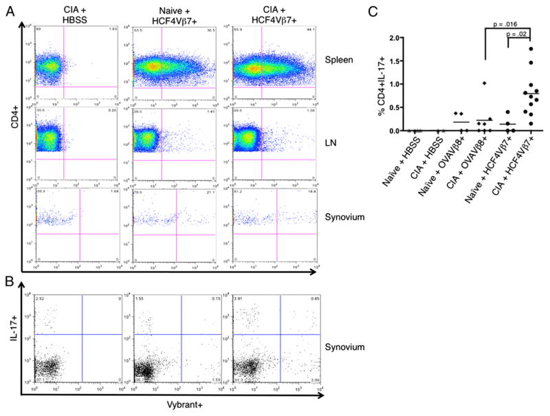
FIGURE 6.
In vivo, IL-17–producing HCF4Vβ7+ CD4 T cells migrate to joints. Naive mice or mice with CIA, as described in Materials and Methods, were injected with 1 × 10⁷ T cells stained with Vybrant at day 24 of the protocol, and spleens, LN, and knee synovium were harvested 12 d later. The negative control for these studies was a mouse with CIA injected with HBSS (CIA + HBSS). (A) Plots show representative examples of expression of Vybrant-stained HCF4Vβ7+ T cells analyzed by flow cytometry in naive mice or mice immunized with bCII. Cells from the spleen, LN, and synovium are displayed. (B) Inflammatory cytokine expression was analyzed by intracellular staining, following incubation with PMA-ionomycin and Golgi plug. Plots show representative examples of IL-17 expression by Vybrant-stained HCF4Vβ7+ T cells from knee synovium in naive mice or mice immunized with bCII. (C) The percentage of Vybrant+IL-17+ CD4 T cells in the synovium (upper right quadrant in B) was quantified for each group of mice, as well as in naive mice and mice with CIA injected with the OVAVβ8 line. Each bar indicates the mean, and the relevant statistical significance is displayed. n = 2 mice injected with HBSS, n = 11 mice injected with the HCF4Vβ7+ line, and n = 7 mice injected with the OVAVβ8+ line.

