Figure 3.
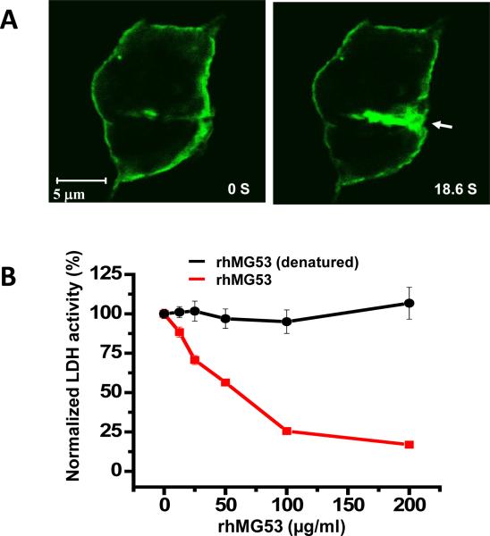
In vitro assay with MG53 protection against injury to cultured RLE cells. A. RLE cell transfected with GFP-MG53 show rapid translocation of GFP-MG53 containing intracellular vesicles toward the acute plasma membrane injury site following penetration of a microelectrode. Left panel – cell image taken immediately after injury, right pane – image taken 18.6 s after injury. Arrow shows the microelectrode injury site. Visualization of live cell imaging of the GFP-MG53 movement process can be found in Supplemental Movie 1. B. RLE cells were treated with external rhMG53 or boiled rhMG53 (denatured control protein) in the cell culture medium and then exposed to mechanical membrane damage by glass beads. Membrane damage is measured by LDH release from cells. rhMG53 reduced LDH release due to mechanical damage and this protective effect is dose dependent, n=9-12 for each data point (mean ± SEM).

