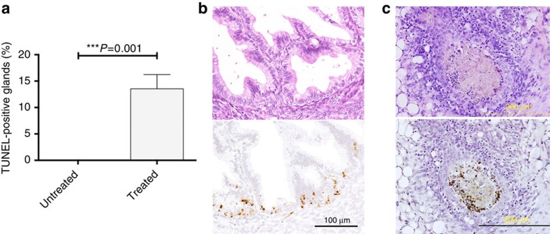
Figure 6. Induction of apoptosis in baboon endometriosis tissue in vivo.
(a) Quantitative TUNEL analysis. Shown is the percentage of TUNEL-positive endometrial glands in tissues from three control untreated baboons compared with three animals treated with a mixture of dKLAK-z13 and HLAH-z13 peptides (see Methods). The number of glands counted and the number of TUNEL-positive glands (indicated in parentheses) were 5 (0), 8 (0) and 18 (0) for untreated and 8 (1), 18 (2) and 26 (3) for treated. Percentages of TUNEL-positive glands from untreated and treated animals were 0.00%±0.00 (n=3) and 13.53%±1.56 (n=3), respectively. (b) TUNEL assay of baboon endometriosis. Upper panels: hematoxylin and eosin-stained images. Lower panels: TUNEL assay of corresponding areas. Left: an endometrial gland in the ovary. Scale bar, 100 μm. Right: TUNEL-positive cell debris in an endometrial gland in the omentum. Scale bar, 200 μm.

