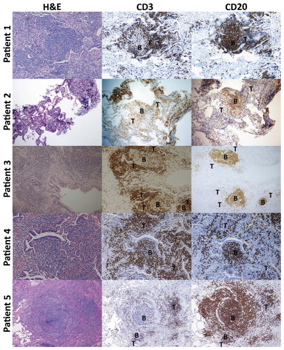
FIG 2.
Pulmonary lymphocytes are organized into B- and T-cell zones in patients with CVID-associated PLH. Hematoxylin-eosin and immunohistochemical staining for CD3 (T cells) and CD20 (B cells) is shown for 5 patients. Areas of B- and T-cell predominance are labeled as B and T, respectively. Magnification of images is ×40.

