
Figure 1.
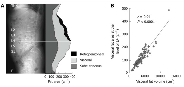
Correlation between visceral fat volume and visceral fat area measured by multislice computed tomography. A: Abdominal fat distribution; B; Correlation between abdominal visceral fat volume and visceral fat area from a single slice at the level of L4. Number = 75 (males 55, females 20); age: 53 ± 1 yr (range, 18-81 yr), body mass index: 25.4 ± 3.9 kg/cm2 (18.9-38.8 kg/cm2). Slice thickness was 10 mm, and images were obtained from Th 8/9 to the pubis. Fat area measurement application: Advanced area calculation, GE Healthcare Co.