
Figure 4.
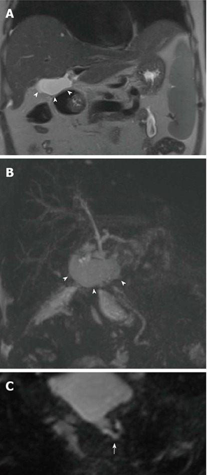
Bile leakage in a 54-year-old male subject transplanted for hepatitis C virus-related cirrhosis. Perihilar biloma shown by arrowheads on coronal T2-weighted HASTE image (A) and paracoronal MIP reconstruction from 3D MRC (B). Thin communication between the anastomotic site and fluid collection is visible on the axially-reformatted 3D source image (arrow in C). MRC: Magnetic resonance cholangiography; MIP: Maximum intensity projection.