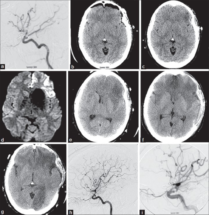
Figure 1.

Tracing the month long course of life-threatening vasculitis after aneurysm clipping in a 33-year-old woman. Initial clipping. (a) After coil embolization for rupture of a right internal carotid artery (ICA) aneurysm, an unruptured left posterior communicating artery (PComA) aneurysm was incidentally detected and then treated by clipping; this intraoperative angiogram demonstrated flow in PComA and fetal posterior cerebral artery (PCA) after clipping and no residual aneurysm filling. (b) Postoperative CT confirms clipping was successful. Readmission and reclipping. Eleven days later (day 1), patient returns to the emergency department where head CT (c) and MRI (d) showed acute infarction in the orbitofrontal and left frontal opercular cortical regions. CTs during hospital days 11–22. Day 11 (e), progressive mass effect and infarction; day 12 (f), after surgical decompression, evolution of new bifrontal infarctions and day 16 (g) demonstrating new left PCA infarct with worsening of bilateral frontal infarctions. Day 17 (h), repeat angiogram shows high-grade stenosis and near-complete occlusion of the left PCA at the P1-2 junction; mild short segment stenosis involves several cortical branch vessels of left MCA. Day 22 (i), intraoperative angiogram after nickel-containing clip removal was replaced with titanium clip
