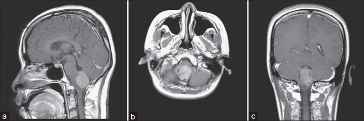
Figure 1.

Preoperative brain MRI. T1-weighted postcontrast image, sagittal (a), axial (b), coronal (c) views revealed a heterogeneous enhancing mass lesion with several foci of cystic change over inferior fourth ventricle, about 3.2 × 3 × 2.6 cm, and extension to foramen magnum, causing mild dilatation of fourth ventricle
