Figure 3.

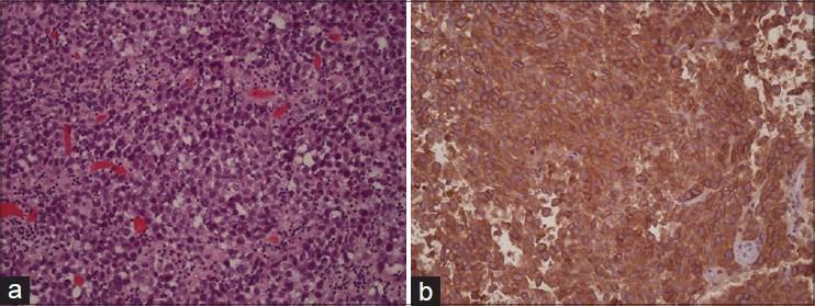
Histology of the tumor. H and E, ×200 (a) shows the specimen composed of sheets of large anaplastic cells divided by delicate fibrovascular septae with small lymphocytes, mitosis and necrosis. CD117 stain (b) shows the positive staining

Histology of the tumor. H and E, ×200 (a) shows the specimen composed of sheets of large anaplastic cells divided by delicate fibrovascular septae with small lymphocytes, mitosis and necrosis. CD117 stain (b) shows the positive staining