Figure 5.

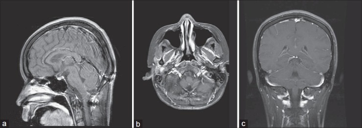
1-year postoperative brain MRI. T1-weighted postcontrast image, sagittal (a), axial (b), coronal (c) views demonstrated neither residual nor recurrent tumor

1-year postoperative brain MRI. T1-weighted postcontrast image, sagittal (a), axial (b), coronal (c) views demonstrated neither residual nor recurrent tumor