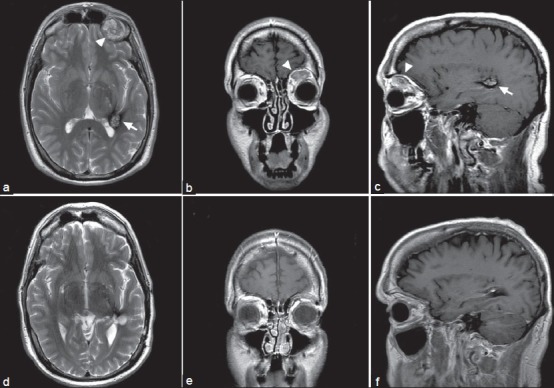
Figure 1.

MRI of the CCM and OCH pre- and post-resection (at 3-year follow-up). (a) Axial T2 MRI showing left temporal heterogeneous popcorn lesion (arrow) extending into the atrium of the left occipital horn of the lateral ventricle consistent with a CCM. A left orbital lesion consistent with an orbital hemangioma is also visualized (arrowhead). (b) Coronal T2 image showing OCH. (c) Sagittal T1 image showing the CCM and OCH before resection. (d-f) Axial T2, coronal T2, and sagittal T1 MRIs post-resection demonstrating complete resection of the CCM and OCH
