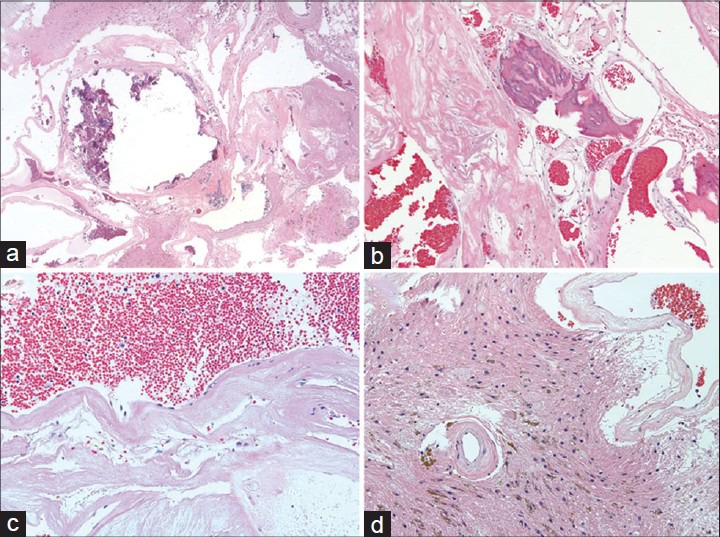
Figure 3.

(a, b) CCM comprised cavernous, endothelium-lined vascular sinusoids with foci of calcification (a) and ossification. (b) Little intervening brain tissue between the cavernous vessels was noted. (c) High-power view of the vascular walls of the CCM demonstrates delicate mural hyalinization, scattered extravasated erythrocytes and hemosiderin, and scant inflammation. (d) Brain parenchyma at the periphery of the lesion showing typical hemosiderin deposits, macrophages, axonal spheroids, and gliosis
