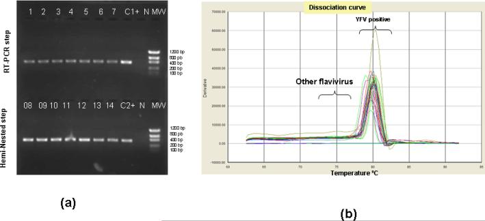
Figure 1.
Detection of YFV sequences in extracts of infected Vero cells. (a) Size fractionated, ethidium bromide labeled product (4μL) of RT-(lanes 1 to 7) and RT-hemi- nested-PCR (lanes 8 to 14) steps (1% agarose gel). (b) the dissociation curve for the SYBR® qRT-PCR showing the melting temperature for positive YFV genome detection ranging from 79°C to 81°C (mean of 80°C). “C+1 and C+2” stand for positive controls used during the RT-PCR and Hemi-nested-PCR steps, respectively. “NC” stands for negative control (RNA extracted from supernatant of uninfected Vero cells) and “MW” stands for molecular weight marker (100bp DNA ladder, Invitrogen, Carlsbad, CA, USA).

