
Figure 3. A chronic cervical hemi-contusion model that recapitulates human pathology.
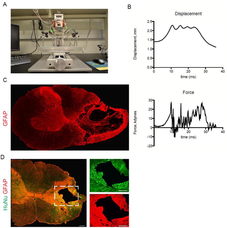
(A) Fourth generation of the OSU injury device. (B) Contusion injury data: representative displacement and force tracings from one injury. (C) Four weeks after injury, GFAP staining reveals substantial cavitation and reactive gliosis on the side of contusion. (D) Eight weeks after transplant, hiPSC-NPCs have survived and integrated in the chronic contusion model. Most cells remain close to the site of injection (i.e. near the gray-white border of the dorsolateral funiculus and juxtaposed to the lesion cavity), but many migrated throughout the ipsilateral white and gray matter.