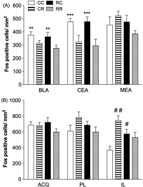
Figure 3. Fos-IR labeling in amygdala and mPFC.
(A) Groups that were placed in a novel context but not restrained on day 2 exhibited elevated Fos expression in BLA and CEA compared to those that were restrained (**p < 0.01 vs. CR, RR in BLA; ***p = 0.001 vs. CR, RR in CEA; n=5-8/group).(B) Restraint and context conditions altered Fos labeling within the IL without significantly affecting the PL or the ACG. Specifically, rats placed in the previous day's restraint context (RC) but not restrained showed elevated Fos-IR labeling within the IL compared to those that were never restrained (#p < 0.05 vs. CC). Rats that were acutely restrained (CR) also exhibited greater Fos-IR labeling compared to those that were never restrained (##p < 0.01 vs. CC; n=3-8/group).

