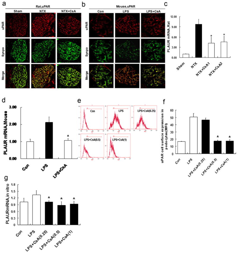
Fig. 2.
CsA inhibits podocyte uPAR expression in vivo and in vitro. a Double immunofluorescence staining for uPAR (red) and the podocyte marker synaptopodin (synpo, green) in glomeruli from NTX rats treated with or without CsA, as evaluated by confocal microscopy. The podocytes of untreated NTX rats showed an increased expression of uPAR protein. CsA substantially inhibited uPAR induction. b Same as in Fig. 2a, but now in the LPS mice. LPS substantially enhanced podocyte uPAR induction. CsA significantly inhibited uPAR induction. c Quantitative real-time RT-PCR was performed on kidney glomerulus isolated from NTX rats. PLAUR mRNA (encoding uPAR) was upregulated in untreated NTX rats. CsA inhibited PLAUR mRNA expression (p<0.01). d Same as in Fig. 2c, but now in the LPS mice. Untreated LPS mice showed an increased PLAUR mRNA expression. CsA inhibited PLAUR mRNA expression (p<0.01). e, f Flow cytometry for uPAR cell surface expression showed the development of a high uPAR population after LPS treatment. CsA (0.25–1 μg/ml) reduced uPAR cell surface expression (p<0.01). g Quantitative real-time RT-PCR was performed on cultured differentiated podocytes. PLAUR mRNA expression in LPS-treated podocytes was upregulated. Treatment with CsA (0.25–1 μg/ml) inhibited PLAUR mRNA expression (p <0.01). All values are expressed as the means±SD. *p<0.01 versus untreated NTX rats or LPS mice or podocytes treated with LPS only

