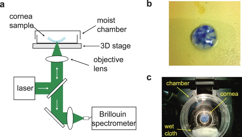
Figure 1.
Experimental Setup. (a) Schematic of the measurement setup. (b) Representative picture of a typical corneal central “button” sample. The blue ink marks helped identify the position of the cone (see also insets in Fig. 3). (c) The sample is placed face down on a glass-bottom culture dish with a wet cloth to keep the moist environment during measurements.

