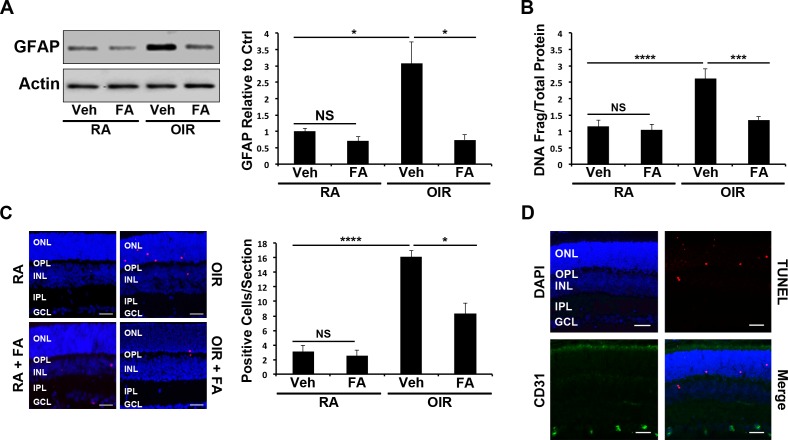
Figure 1.
Cytoprotective effect of Feno-FA (FA) in the retinas of WT OIR mice. (A) At P17, retinal GFAP was measured by Western blotting in room air (RA) and OIR mice treated with Vehicle control (Veh) or FA, semiquantified by densitometry, normalized by β-actin levels, and expressed as the ratio to that in the RA control (n = 6). (B) Retinal DNA fragmentation in P17 mouse retinas treated as above was quantified using a cell death ELISA and normalized to total protein concentration (n ≥ 5). (C) Apoptotic cells in retinal sections from mice treated as above were labeled with TUNEL staining (red), and the nuclei counterstained with DAPI (blue). TUNEL-positive cells were counted in retinal sections (n ≥ 6); scale bars: 10 μm. (D) Retinal sections were double stained with anti-CD31 antibody (green) and TUNEL (red), and the nuclei were counterstained with DAPI (blue) (n ≥ 3). Scale bars: 10 μm. All values are mean ± SEM; *P ≤ 0.05, ***P ≤ 0.001, ****P ≤ 0.0001. OPL, outer plexiform layer; IPL, inner plexiform layer; GCL, ganglion cell layer.

