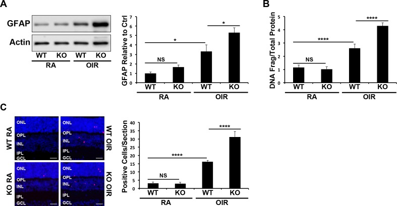
Figure 3.
Increased OIR-induced retinal cell death and glial activation by PPARα ablation. (A) Retinal GFAP expression in P17 RA and OIR WT and PPARα−/− mice was measured by Western blot analysis, normalized by β-actin levels, and expressed as the ratio to that in WT RA control (n = 6). (B) Retinal DNA fragmentation in mice treated as above was measured by cell death ELISA and normalized by total retinal protein levels (n ≥ 5). (C) Apoptotic cells in retinal sections from mice treated as above were labeled with TUNEL staining (red), and nuclei were counterstained with DAPI (blue). TUNEL-positive cells were counted in retinal sections (n ≥ 6); scale bars: 10 μm. All values are mean ± SEM. *P ≤ 0.05, ****P ≤ 0.0001.

