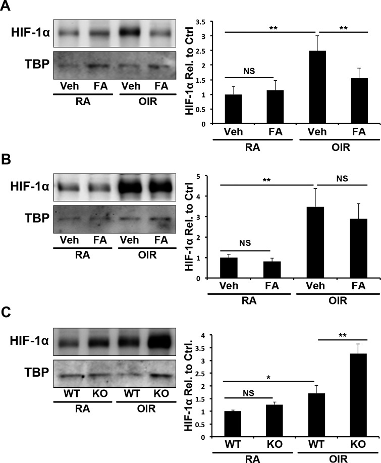
Figure 7.
Inihibition of HIf-1α by PPARα in OIR. (A) In P17 WT RA and OIR mice, retinal levels of nuclear HIF-1α were measured in Veh and FA-treated mice by Western blot analysis, normalized by TBP levels and expressed as the ratio to vehicle-treated RA control (N ≥ 5). (B) In P17 PPARα−/− RA and OIR mice, retinal nuclear HIF-1α levels were measured as above (N ≥ 5). (C) In WT and PPARα−/− RA and OIR mice, retinal nuclear HIF-1α levels were measured as described above and normalized to RA WT mice (N ≥ 5). All values are mean ± SEM. *P ≤ 0.05; **P ≤ 0.001.

