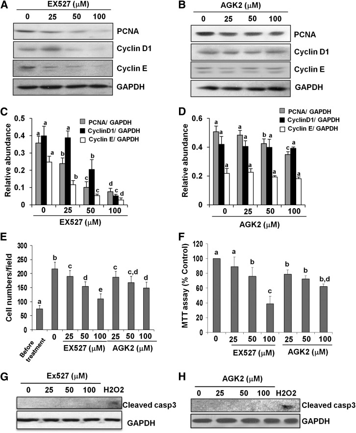
Fig. 4.
Effects of SIRT1 and SIRT2 inhibitors on renal fibroblast proliferation. NRK-49F cells were cultured in medium with 5% fetal bovine serum and treated with EX527 (0–100 μM) and AGK2 (0–100 μM) for 36 hours (A–F). Then, cell lysates were prepared and subjected to immunoblot analysis with antibodies for PCNA, cyclin D1, cyclin E, or glyceraldehyde-3-phosphate dehydrogenase (GAPDH; A and B). Representative immunoblots from three experiments are shown. The levels of PCNA, cyclin D1, and cyclin E were quantified by densitometry and normalized with GAPDH (C and D). NRK-49F cells were treated with the indicated concentration of EX527 and AGK2 for 36 hours, cells were randomly photographed in bright field (200×), and cell proliferation was measured by cell counting (E) or the MTT assay (F). To measure cell death, cultured NRK-49F cells were exposed to the same concentrations (0–100 μM) of EX527 or AGK2 for 48 hours or treated with 1 mM H2O2 for 3 hours as positive control. Cell lysates were subjected to immunoblot analysis for cleaved caspase-3 and GAPDH (G and H). Values are the means ± S.D. of three independent experiments. Bars with different letters (a–e) are significantly different from one another (P < 0.01).

