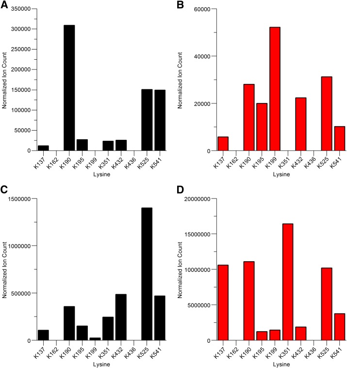
Fig. 5.
Ion-count epitope profiles of HSA modified at lysine residues by reactions with diclofenac 1-β AG in vitro. (A) Acylation adducts at 0.5 hour. (B) Glycation adducts at 0.5 hour. (C) Acylation adducts at 16 hours. (D) Glycation adducts at 16 hours. Data represent relative ion intensities of lysine-modified peptides detected during LC-MS/MS analyses of tryptic digests. Each relative ion intensity was derived from the area under the curve for the relevant extracted ion chromatogram by normalization to the total ion count of the sample. Synthetic AG was incubated with HSA (AG:HSA molar ratio, 50:1) at pH 7.4 and 37°C. Acylated Lys351 was detected in these analyses, but this modification of Lys351 and also modifications of Lys162 and Lys436 were not detected consistently (Table 2). Lys436 was modified detectably in patient N08.

