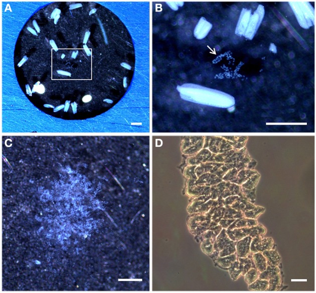
Figure 3.

Stereomicroscopy and light microscopy images of rice anthers and isolated meiocytes. (A) Stereomicroscopy observations of rice anthers suspended in a solution PBS and 1% proteinases inhibitors on a multi-well slide. (B) Inset in (A) shows the meiocytes bag (arrowed) from a sectioned rice anther. (C) Rice isolated meiocytes under the stereomicroscope. (D) Light microscopy observations of rice isolated meiocytes in a Neubauer chamber. Bar represents 500 μm in (A,B), and 20 μm in (C,D).
