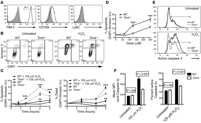
Figure 5. CD169+ peritoneal macrophages are resistant to H2O2-induced apoptosis in Tim4–/– relative to WT mice.
(A) Histograms of gated WT or Tim4–/– DAPI–CD11b+F4/80+ peritoneal macrophages revealed TIM-4 and CD169 expression, but not Ly6C expression. Shaded histograms represent indicated controls. MFI is noted parenthetically. (B–D) Gated WT or Tim4–/– CD11b+F4/80+ peritoneal macrophages incubated in the presence or absence of H2O2 (125 μM unless otherwise indicated) were analyzed for DAPI and annexin V binding by flow cytometry. (B) Representative flow cytometry plots at 3 hours after treatment. (C) Summary graphs from 1 of 3 independent experiments, showing lower apoptosis (DAPI–annexin V+) and cell death (DAPI+annexin V+) in Tim4–/– versus WT CD11b+F4/80+ macrophages. *P < 0.05, **P < 0.01, ***P < 0.001, ANOVA (P < 0.0001) with Bonferroni post-test. (D) Dose-response curve showing less apoptosis (DAPI–annexin V+) in Tim4–/– versus WT CD11b+F4/80+ macrophages at 3 hours. ***P < 0.001, ANOVA (P < 0.01) with Bonferroni post-test. (E and F) Gated CD11b+F4/80+ macrophages treated with 125 μM H2O2 for 3 hours were permeabilized and stained for intracellular active caspase-3 in triplicate. Shown are representative histograms (E) and summary graphs showing active caspase-3 MFI and frequency of active caspase-3+ macrophages from 1 of 2 independent experiments (F). Active caspase-3 levels in gated Tim4–/– macrophages was reduced compared with WT macrophages. P values were determined by ANOVA (P < 0.01) with Bonferroni post-test. All graphs depict mean ± SEM from a representative experiment performed in triplicate.

