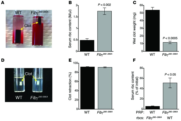
Figure 2. rbc are extruded from Fibγ390–396A clots during clot retraction, resulting in decreased clot weight.
After clot formation, platelet contractile force retracts clots away from siliconized tube walls, leaving the clots surrounded by serum. (A) Image of fully retracted WT and Fibγ390–396A clots 90 minutes after initiation of clot formation by tissue factor and CaCl2. (B) Number of rbc present in the serum following clot retraction (n = 3). (C) Wet clot weights following clot retraction (n = 3). (D) Image of fully retracted PRP clots 90 minutes after initiation of clot formation. (E) Percentage retraction of PRP clots (n = 3). (F) Serum rbc content of retracted clots containing WT PRP with Fibγ390–396A rbc and vice versa (n = 3). Data represent mean ± SEM.

