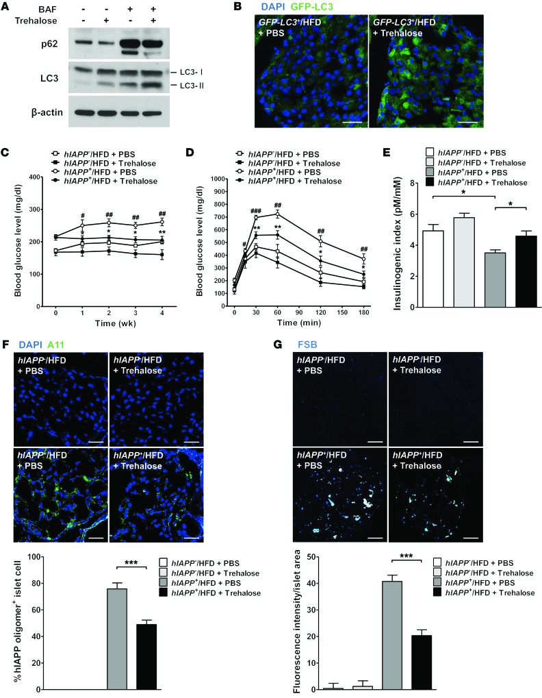
Figure 6. Effects of autophagy enhancer on the glucose profile of hIAPP+ mice.
(A) Primary mouse islet cells were treated with 100 mM trehalose for 24 hours with or without 100 nM bafilomycin pretreatment to block lysosomal steps of autophagy. Cell lysates were subjected to Western blot analysis using anti-LC3 or anti-p62 Ab. (B) Trehalose (2 g/kg) was administered i.p. to 12-week-old GFP-LC3+ mice on HFD for 2 weeks, and frozen pancreas sections were prepared for confocal microscopy to examine LC3 puncta. Scale bar: 50 μm. (C) Trehalose (2 g/kg) was administered i.p. to 16- to 20-week-old hIAPP+ mice on HFD, and nonfasting blood glucose levels were monitored (n = 5). (D) IPGTT was performed after 4 weeks of trehalose administration to hIAPP+ mice on HFD (n = 5). (E) The insulinogenic index was calculated after 4 weeks of trehalose administration to hIAPP+ mice on HFD (n = 3). (F) After immunofluorescent staining using A11 Ab, the percentage of A11-stained cells among total DAPI+ islet cells was determined by confocal microscopy. Representative pictures are shown. Scale bar: 50 μm. (G) After FSB staining, mean fluorescence intensity per islet area was determined using the NIS-Elements AR 3.0 software (Nikon). Representative pictures are shown. Scale bar: 100 μm. *P < 0.05, **P < 0.01, ***P < 0.001; #P < 0.05, ##P < 0.01, ###P < 0.001. (*, comparison between hIAPP+/HFD + trehalose and hIAPP+/HFD + PBS groups; #, comparison between hIAPP+/HFD + PBS and hIAPP–/HFD + PBS groups in C and D.)

