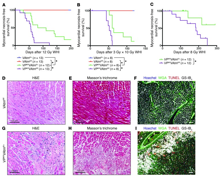
Figure 4. Deletion of Atm in p53 WT endothelial cells does not sensitize mice to radiation-induced myocardial necrosis.
(A) Kaplan-Meier plots of myocardial necrosis–free survival for VAtmfl/+, VAtmfl/fl, VPfl/flAtmfl/+, and VPfl/flAtmfl/fl mice after 12 Gy whole-heart irradiation. (B) Kaplan-Meier plots of myocardial necrosis–free survival for VAtmfl/+, VAtmfl/fl, VPfl/flAtmfl/+, and VPfl/flAtmfl/fl mice after whole-heart irradiation with 10 daily fractions of 3 Gy. 1 VPfl/flAtmfl/fl mouse died prior to finishing irradiation and was censored. (C) Kaplan-Meier plots of myocardial necrosis–free survival for VPfl/flAtmfl/+ and VPfl/flAtmfl/fl mice after 8 Gy whole-heart irradiation. Mice of both genotypes were censored due to development of thymic lymphomas prior to heart disease. (D–I) Representative sections of the myocardium of (D–F) a VAtmfl/fl mouse 469 days after whole-heart irradiation and of (G–I) a VPfl/flAtmfl/+ mouse 56 days after whole-heart irradiation, subjected to staining with H&E (D and G) or Masson trichrome (E and H) or immunofluorescence for WGA, TUNEL, and GS-IB4 (F and I). *P < 0.05. Scale bars: 100 μm (D–I).

