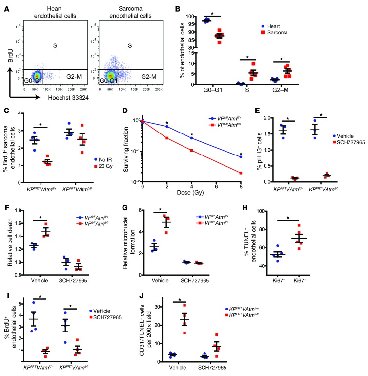
Figure 6. Atm deletion sensitizes proliferating endothelial cells to radiation.
(A and B) Flow cytometry analysis (A) and quantification of cell cycle phase (B) in heart and sarcoma endothelial cells from KPFRT mice (n = 5). (C) Flow cytometry quantification of BrdU incorporation into tumor endothelial cells from KPFRTVAtmfl/+ and KPFRTVAtmfl/fl mice 1 hour after irradiation with 20 Gy or in unirradiated controls (n = 4 per group). (D) Clonogenic assay of primary cardiac endothelial cells from VPfl/flAtmfl/+ and VPfl/flAtmfl/fl mice (n = 3 independent experiments). (E) Flow cytometry quantification of phosphorylated histone H3 (pHH3) for primary cardiac endothelial cells from VPfl/flAtmfl/+ and VPfl/flAtmfl/fl mice treated with DMSO vehicle or 500 nM SCH727965 for 24 hours. (F and G) Cell death (F) and micronuclei formation (G), 24 hours after irradiation of primary cardiac endothelial cells from VPfl/flAtmfl/+ and VPfl/flAtmfl/fl mice treated with DMSO or 500 nM SCH727965 immediately before irradiation with 12 Gy (n = 3 independent experiments). Data are expressed relative to unirradiated cells of the same genotype and drug treatment. (H) Quantification of TUNEL staining in Ki67+ and Ki67– endothelial cells (CD31+) from tumors in KPFRTVAtmfl/fl mice 24 hours after irradiation with 20 Gy (n = 5). (I) Flow cytometry quantification of BrdU incorporation into sarcoma endothelial cells from KPFRTVAtmfl/+ and KPFRTVAtmfl/fl mice 24 hours after injection with vehicle or 40 mg/kg SCH727965 (n = 4 per group). (J) Quantification of CD31+TUNEL+ cells in sarcomas from KPFRTVAtmfl/+ and KPFRTVAtmfl/fl mice 24 hours after treatment with vehicle or SCH727965 immediately before irradiation with 20 Gy (n = 4 per group). All data are mean ± SEM. *P < 0.05.

