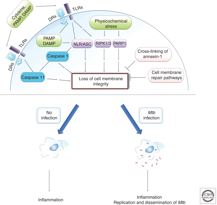
Figure 2.
Necroptosis and pyroptosis both lead to the loss of cell membrane integrity. Necroptosis can be initiated by extracellular ligands interacting with their cognate cell surface receptors (DRs, death receptors; TLR, Toll-like receptors) or by varied intracellular stress signals (physicochemical stress). One pathway involves activation of RIPK1 and RIPK3, which leads to mitochondrial malfunction. Intracellular stress can also activate PARP1, which can signal for mitochondrial dysfunction potentially involving RIPK1 (sometimes referred to as the parthanatos death pathway). The inflammasome (NLR/ASC) may signal directly for necroptosis induction or indirectly via caspase 1 leading to pyroptosis, which also results in cell lysis but induces DNA degradation similar to apoptotic cell death. Caspase 11 can signal directly for a pyroptosis-like cell death or indirectly via NLR/ASC to activate caspase 1–dependent pyroptosis signaling. The cell may attempt to inhibit cell membrane lysis either by recruitment of lysosomal membranes to the cell surface to repair the cell membrane or by cross-linking annexin-1 on the cell surface and thus leading to formation of a stable apoptotic envelope. Necrotic cell death is inflammatory as it leads to spilling of the intracellular contents into the extracellular milieu. Mtb has been associated with both the induction of necrosis pathways and the inhibition of the inhibitory mechanisms. Programmed necrosis of Mtb-infected host cells leads to bacterial escape, whereas the associated inflammatory response recruits new host cells for extracellular Mtb to infect and replicate in, thus leading to dissemination of infection.

