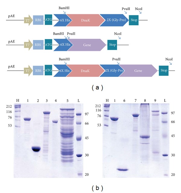
Figure 1.

(a) Schematic representation of the expression cassettes. The genetically fused genes were obtained from individually cloned genes in pAE vector, and, then, surface protein genes were digested at PvuII/NcoI restriction sites and ligated in the same sites into pAE-DnaK construct. Depicted are T7 phage RNA polymerase promoter, ribosome-binding site (RBS), ATG start códon, 6X Histidine tag, restriction cloning sites, and the 2X (Gly-Pro) flexible hinge. (b) Analysis of purified recombinant proteins by SDS-PAGE. Purified recombinant protein eluted from Ni+2-charged Sepharose column with 1 M imidazole are visualized by Coomassie blue staining. Lane H (HMW) and L (LMW): high and low molecular mass protein markers; In kDa: lane 1: DnaK (68.9); lane 2: rLIC10494 (25.1); lane 3: DnaK-rLIC10494 (92.5); lane 4: rLIC12730 (75.7); lane 5: DnaK-rLIC12730 (143.1); lane 6: Lsa21 (19.8); lane 7: DnaK-Lsa21 (87.2); lane 8: Lp95 C-terminal (43.6); lane 9: DnaK-Lp95 C-terminal (110.9).
