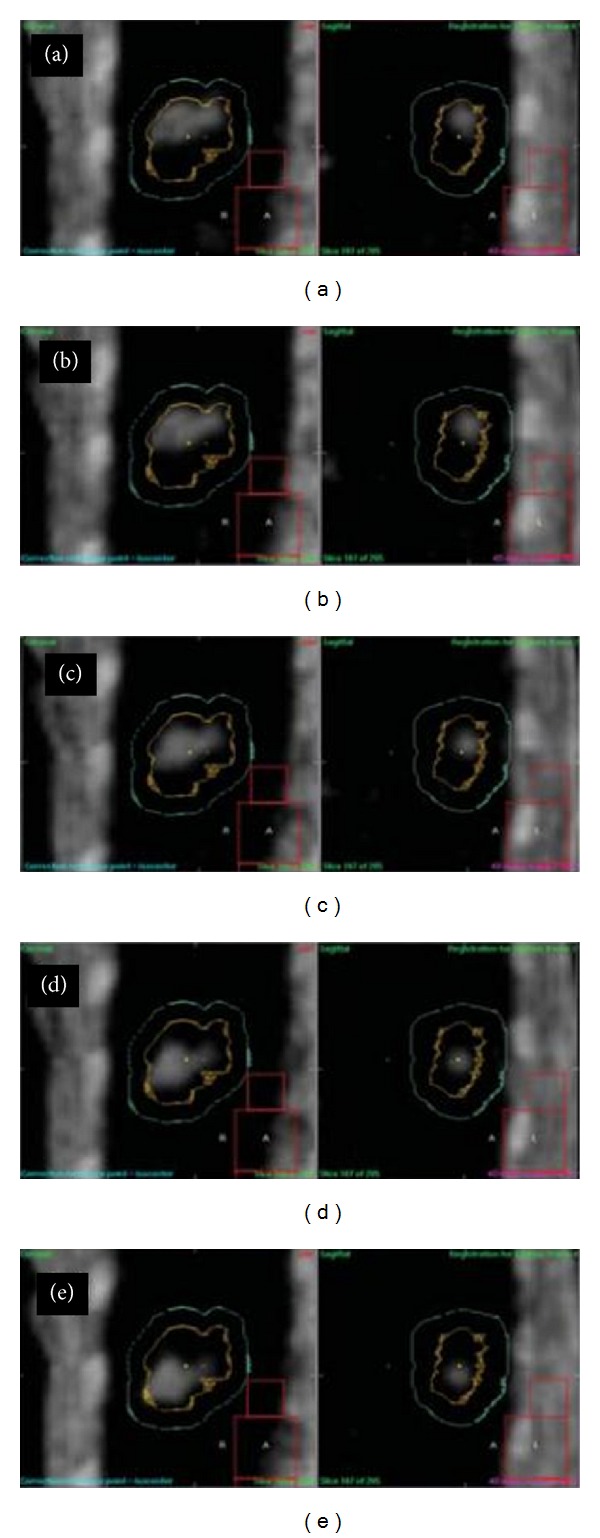
Figure 2.

4D CBCT images on the first day overlaid with PTV (in sky blue) and ITV (in yellow) contours after lung tumor registration for five consecutive respiratory phases covering half a breathing cycle, where the tumor moves from cranial to caudal direction during the half cycle [9].
