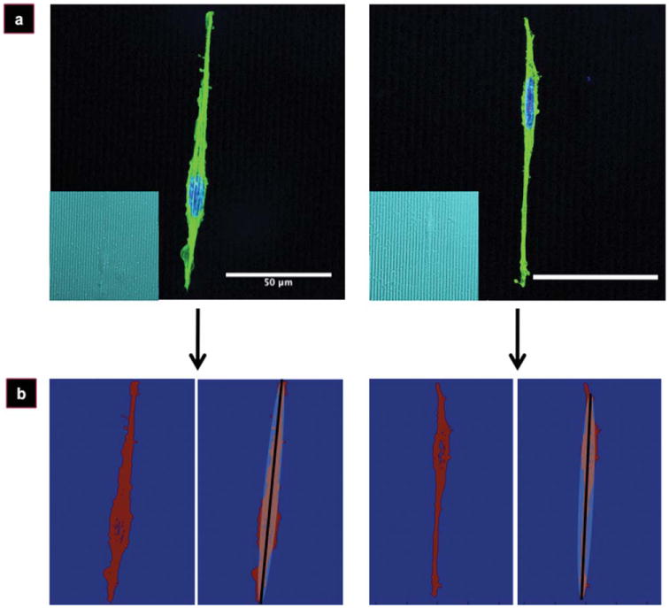
Figure 1.

Imaging of PC-12 cells on silk films. Fluorescent micrographs of representative PC-12 cells (a) on micropatterned silk films (a, inset) used for cellular orientation analysis. Thresholded and discretized images (b, left panels), and the ellipse-fitting result after application of the Gauss–Newton algorithm (b, right panels). Cells stained for nucleus (blue) and actin (green). Scale bar, 50 μm.
