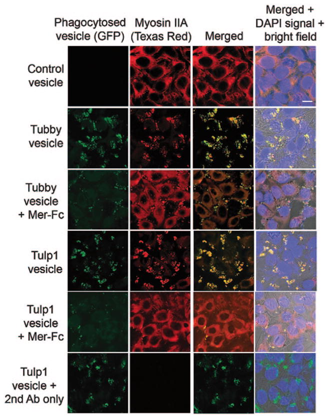
Fig. 64.2.

Tubby- and Tulp1-mediated RPE phagocytosis was MerTK-dependent. mGFP-labeled plasma membrane vesicles were prepared from Neuro-2a cells expressing tubby or Tulp1, and incubated with ARPE19 cells in the presence or absence of excess Mer-Fc (MerTK extracellular domain covalently fused to human IgG1 Fc domain, R&D Systems) for phagocytosis assay as in Fig. 64.1. After washing, ARPE19 cells were fixed, permeabilized, detected with anti-non-muscle myosin II (NMMII) A heavy chain antibodies, detected with Texas red-labeled secondary antibodies and analyzed by confocal microscopy. Fluorescence signals for mGFP and NMMII-A were co-localized. Bar = 10 μm. (Caberoy et al. 2010 [8] with permission from EMBO J)
