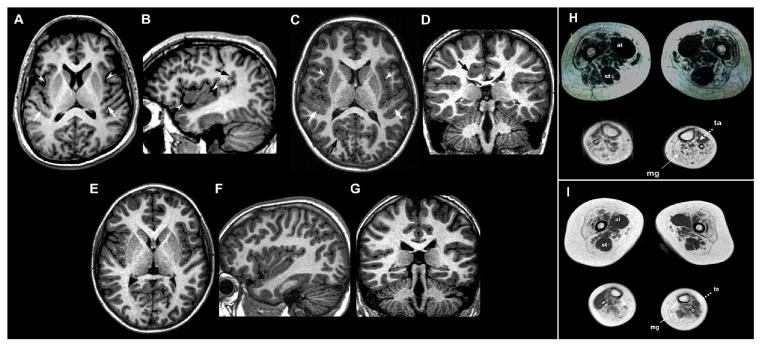
Figure 1.

Brain MRI examinations of Patient 1 (A–B) and Patient 2 (C–D), with corresponding normal control images (E–G) and muscle MRI examination of Patient 1 (H) and Patient 2 (I) at thigh and calf level
Axial A) and right parasagittal B) 3D SPGR T1-weighted images show abnormal gyration and delicate PMG-like cortical malformation involving the insulae (white arrowheads) and Heschle gyri (white arrows) with abnormal vertical orientation of the posterior part of sylvian fissure (black arrow). C) Axial 3D TFE T1-weighted image reveals bilateral perisylvian PMG-like cortical malformation involving the insulae (white arrowheads) and Heschle gyri (white arrows). Note an additional area of abnormal cortical gyration with infolding (black arrows) in the right occipital lobe. D) Coronal 3D TFE T1-weighted image depicts another area of abnormal cortical gyration with infolding (black arrow) in the right posterior cingulum slightly deforming the right lateral ventricle.
In muscle MRIs of lower limbs patients present diffuse atrophy and fat replacement of the thigh muscles which appear more prominent in Patient 2 (I), who accordingly displays the most severe phenotype. A selective similar sparing with compensatory hypertrophy of the adductor longus (al) and semitendinosus (st) muscles is evident to a similar extent in both cases. At leg level, MRI showed severe fat replacement of medial gastrocnemii (mg) with relative preservation of anterior tibialis (ta).
