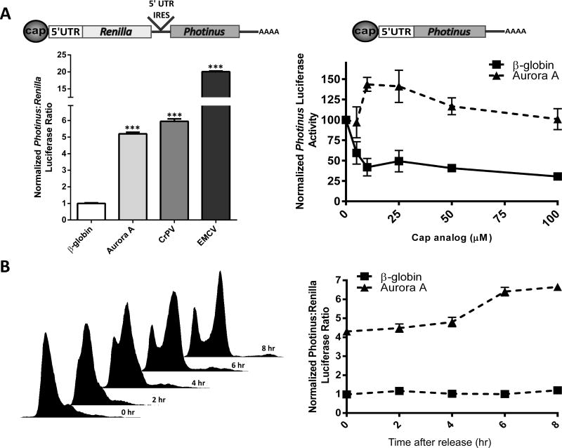
Figure 3.
The 5′ leader of the Aurora A mRNA contains an IRES. (A) Dicistronic luciferase mRNA (left) containing the β-globin, Aurora A, CrPV, and EMCV 5′ leaders or IRES elements were transfected individually into HeLa cells. Luciferase activity is shown as the ratio of Photinus luciferase to Renilla luciferase (P:R) and is normalized to that obtained from the dicistronic mRNA containing the β-globin 5′ leader. n=3 in triplicate ± SD Monocistronic Photinus luciferase mRNA (right) containing the β-globin or Aurora A 5′ leader were translated in rabbit reticulocyte lysate in the presence of increasing concentrations of cap analog. The initial level of Photinus luciferase activity from each monocistronic mRNA was normalized to 100. n=3 ± SD (B) Cell cycle progression following the release of a double thymidine block in HeLa cells was confirmed by flow cytometry analysis (left). HeLa cells transfected with dicistronic luciferase mRNA containing the β-globin and Aurora A 5′ leaders were synchronized in G1/S with a double thymidine block and released. Luciferase activity is shown as the ratio of Photinus luciferase to Renilla luciferase (P:R) and is normalized to that obtained from the dicistronic mRNA containing the β-globin 5′ leader at the 0 time point (right). n=3 in triplicate ± SD, *** = p<0.0001, student’s t test.

