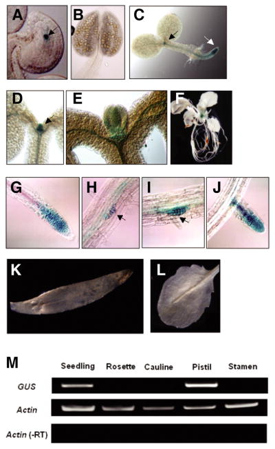
Fig. 2.

Spatiotemporal DME expression during plant development. Transgenic plants containing GUS reporter gene fused with DME promoter region were used to confirm where DME is expressed during plant development. (A) Strong GUS signal was detected in the central cell nucleus (arrow) of the female gametophyte, but not in male gametophytes or stamens (B). (C) In 1-day seedling, GUS was detected primarily in proliferating cells flanking the shoot apical meristem and root apical meristem regions (black and white arrows, respectively). (D–E) Strong GUS signal is restricted in young leaf primordia emerging from the shoot apical meristem in 4-day seedlings. (F) Whole mount view of transgenic plants before bolting. DME promoter is active only in the undifferentiated tissue. (G) GUS was expressed in primary roots adjacent to the meristems. (H–J) GUS was also expressed in secondary roots adjacent to the meristems. (K–L) GUS expression was not detected in mature tissues such as cauline or rosette leaves, respectively. (M) GUS RNA was detected mainly from seedling tissues and pistils that contain undifferentiated proliferating cells and central cell, respectively.
