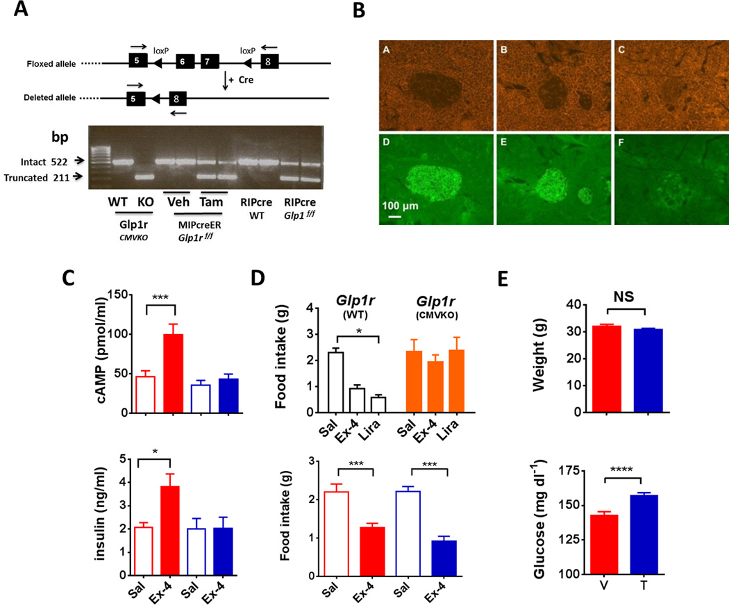
Figure 1. Description and validation of Glp1rf/f and Cre lines.

(A) Upper panel: schematic depicting the location of loxP sites inserted within Glp1r gene and the result of exons 6 and 7 deletion. Lower panel: agarose gel electrophoresis of PCR products from primers designed to generate amplicons spanning exons 6 and 7 in the Glp1r gene; the WT band is 522 bp and the truncated band 211 bp. (B) Pancreatic sections from cross of Cre lines with a “double reporter” (DR). RIPcre × DR (A, D) and MIPcreER × DR treated with tamoxifen (B, E) show diffuse EGFP in the islet under a Cy5 filter, while MIPcreER × DR given vehicle (C, F) have minimal fluorescence in β-cells. (C) Upper panel: cAMP accumulation in isolated islets (40 islets/sample, 8 mice per group, 4 separate isolations) incubated for 15 minutes in media containing IBMX with 10 nM Ex-4 or control (Vehicle red; Tamoxifen blue, *** p ≤ 0.001). (C) Lower panel: insulin concentrations in media from the islet studies described for top panel (Vehicle, red; Tamoxifen, blue, * p ≤ 0.05). (D) Upper panel: no effect of exendin-4 or liraglutide on cumulative 4 hour food intake in Glp1rCMVKO compared with Glp1rWT mice (8 per group); Lower panel: food intake in tamoxifen or vehicle-treated MIPcreER;Glp1rf/f mice (8 per group) in the 6 hours after administration of 2.5 ug Ex-4 IP or saline (*** P ≤ 0.001). (E) Upper panel: body weight in vehicle and tamoxifen treated MIPcreER;Glpr1f/f animals (78 veh and 95 tam treated mice); Lower panel: fasting glucose in vehicle and tamoxifen treated MIPcreER;Glpr1f/f animals (95 veh and 107 tam treated mice; *** P ≤ 0.001). (See also Figure S1 and Figure S2)
