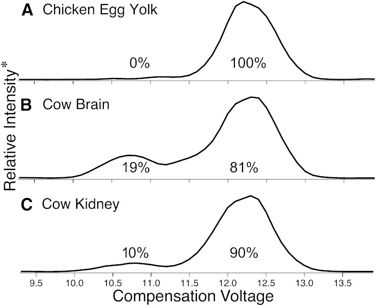
Fig. 4.
Total ionograms resulting from DMS-based separation of [PC (16:0_18:1) + Ag]+ sn-positional isomers formed during positive-mode ESI of lipid extracts from (A) chicken egg yolk; (B) cow brain; and (C) cow kidney. *Relative intensity represents the total ion abundance resulting from CID of m/z 683.2 [M + Ag − 183]+ ions. The listed percentages (± 4 from the error in the slope of the solid trace in Fig. 3) are the relative amounts from peak integration following correction for isobaric contribution from [PC (16:0_18:2) + 109Ag]+ present in each extract (see supplemental Fig. V).

