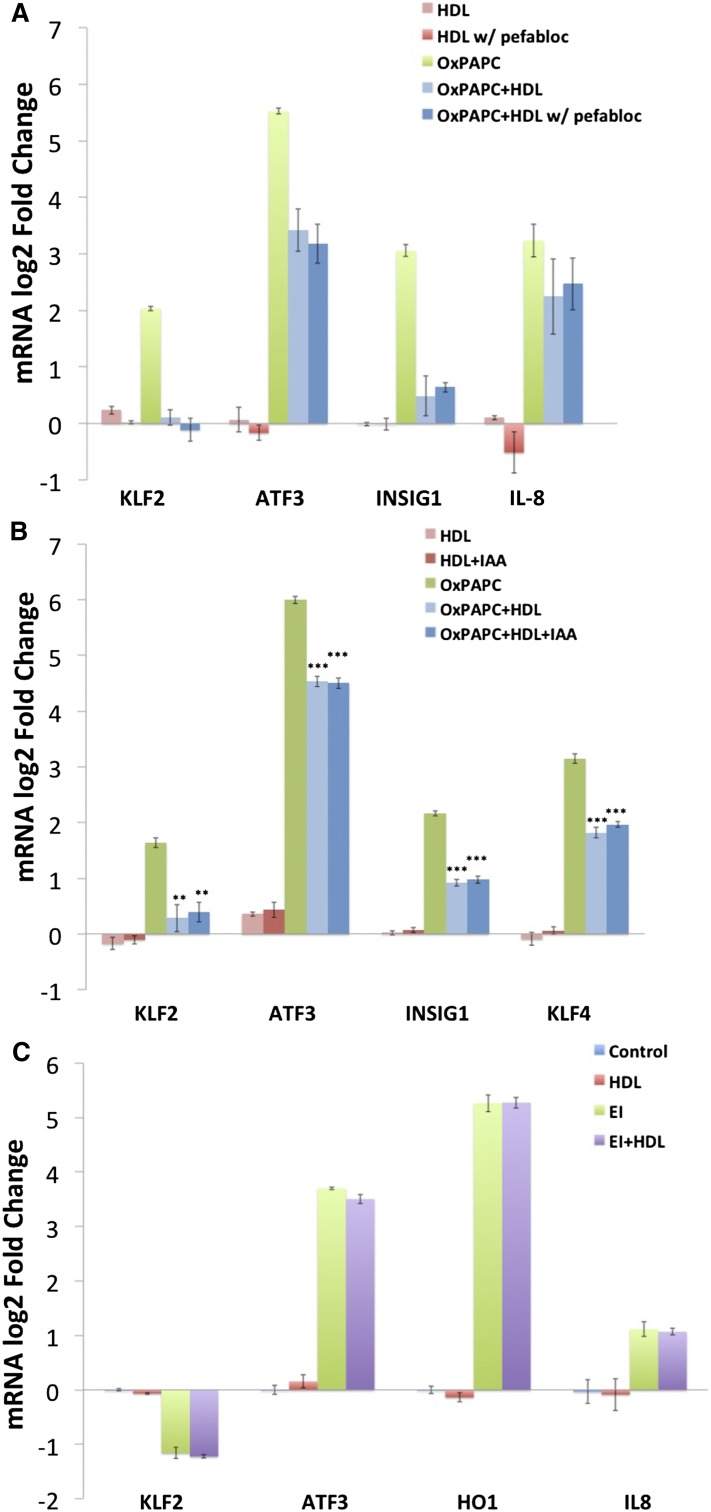
Fig. 6.
The effect of HDL on OxPAPC signaling in HAECs is not dependent on the activity of PAF-AH or direct binding to cysteine residues but is specific to phospholipid components of OxPAPC. HDL (5 mg/ml) was incubated with 1 mM Pefabloc at 37°C for 1 h to inactivate lipoprotein-associated PAF-AH. The HDL was then dialyzed overnight and used in cotreatments with OxPAPC. After the inactivation of PAF-AH, HDL (50 μg/ml) reduced the effects of OxPAPC on markers of inflammatory response (IL-8 and KLF2), ER stress (ATF3), and cholesterol depletion (INSIG1) (A). Shown are the results from one of three representative experiments. Data are presented as average log2-fold change of duplicate samples ± (Max − Min)/2. B: HDL (5 mg/ml) was incubated with 100 mM iodoacetamide at 37°C for 1 h to irreversibly block free thiol residues on HDL. The HDL was then dialyzed overnight and used in HAEC treatments as described in the Materials and Methods. Treatment with iodoacetamide did not affect the ability of HDL to inhibit OxPAPC signaling in HAECs indicating that OxPAPC binding to cysteine residues on HDL is not mediating HDL protection. Data are presented as average log2-fold change of triplicates ± SEM. * P < 0.05, ** P < 0.01, *** P < 0.001 for comparison with OxPAPC log2-fold changes. P values defined by unpaired Student’s t-test. C: HAECs were treated with 1 μg/ml EI in the absence or presence of 50 μg/ml HDL for 4 h as described in the Materials and Methods. Unlike for OxPAPC, HDL cotreatment did not mitigate the effects of EI on the expression of IL-8, KLF2, ATF3, or HO1 as determined by qPCR. Data are presented as average log2-fold change versus control treated samples. All treatments were run in triplicate, and error bars correspond to SEM.

