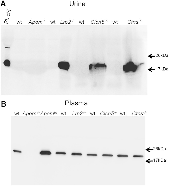
Fig. 4.
Western blotting of apoM in urine (A) and plasma (B) of Apom, Apomtg mice and mice with defective megalin (Lrp2−/−), ClC-5 (Clcn5−/−), and cystinosin (Ctns−/−) and their wt controls. Urine (20 μl) or 11.5 μl of 1/10 diluted plasma were separated by SDS-PAGE and immunoblotted as described in the Methods section. Note the absence of apoM from the urine samples of wt mice, but the presence in urine samples of mice with defective tubular transport (A). By contrast apoM plasma levels appear indistinguishable between wt mice and the different tubular proteinuria models.

