Fig. 3.
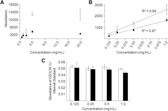
Absolute ion suppression of ganglioside GD3(36:1) ions at m/z 734.9128 as a function of sample concentration measured by negative ionization mode high-resolution (R = 100,000)/accurate MS from modified Svennerholm upper phase retina lipid extracts (open diamonds) and monophasic lipid extracts (filled squares). Serial dilution of each lipid extract was performed from 20 mg/ml (retina tissue extracted/volume of lipid extract) to 0.5 mg/ml (A), and from 1.0 mg/ml to 0.125 mg/ml (B) where the abundance of m/z 734.9128 was observed to follow a linear dependence with retina lipid extract concentration from each extraction method. C: Comparison of relative ion suppression of ganglioside GD3(36:1) at m/z 734.9128, determined as the ratio of the absolute abundance of ganglioside GD3(36:1) to the absolute abundance of a PC(14:0/14:0) internal standard [M+HCO2]− ion at m/z 722.4980, from the modified Svennerholm (open bars) and monophasic (filled bars) lipid extracts at each sample concentration within the linear range shown in (B). Data represent the mean ± standard deviation from n = 3 separate lipid extracts.

