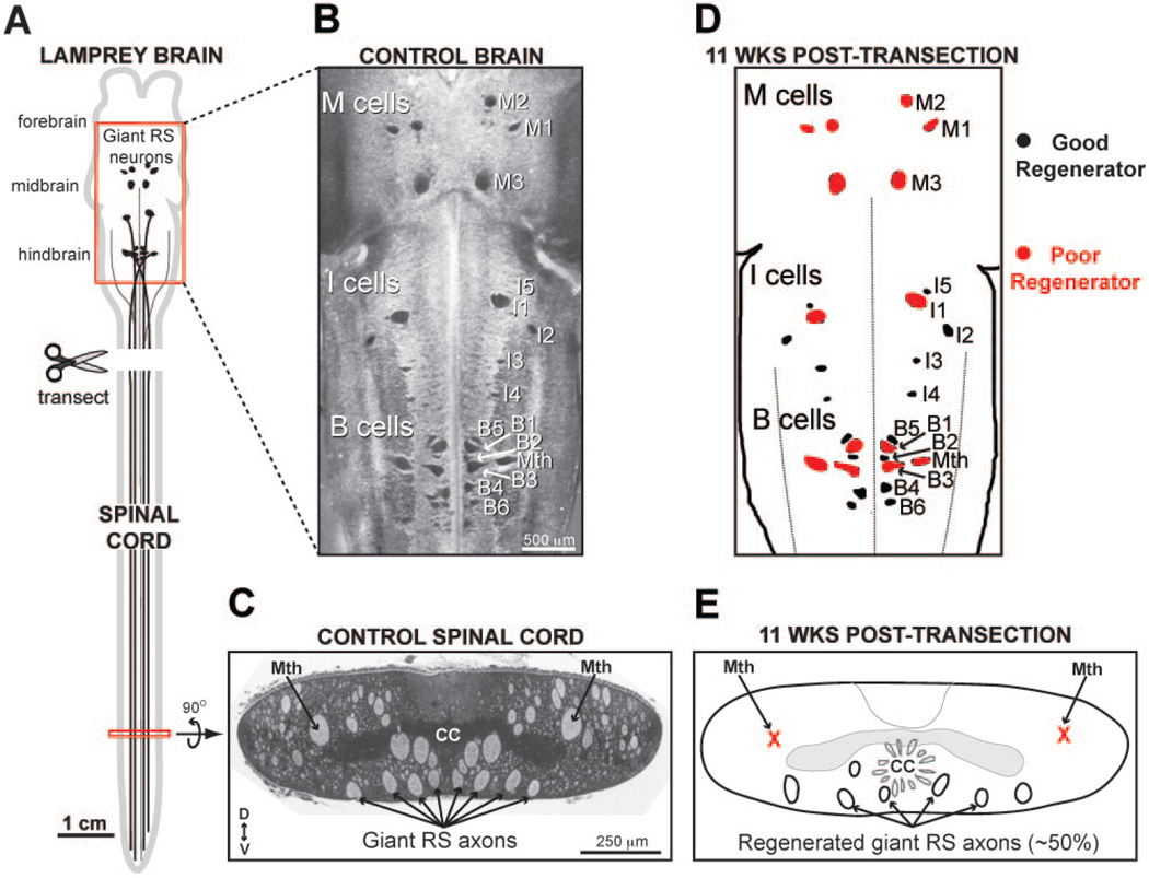
Figure 3.
Regeneration in the lamprey nervous system. (A) Diagram of lamprey CNS. Giant reticulospinal (RS) neurons in midbrain and hindbrain project directly to spinal cord. The typical spinal cord injury paradigm is a complete spinal cord transection, which axotomizes all descending neurons, including giant RS neurons. (B) A Nissl-stained lamprey brain showing the locations of all identified giant RS neurons. Giant RS neurons include the large Müller cells in the mesencephalic (M), isthmic (I), and bulbar (B) brain regions, as well as the Mauthner (Mth) cell. (C) A Nissl-stained cross-section of the lamprey spinal cord showing locations of giant RS axons in the ventromedial tract. The axons of the Mauthner (Mth) neurons are located more dorsolaterally. Dorsal (D) and ventral (V) orientations are indicated. CC = central canal. (D) After transection, some giant RS neurons regenerate reliably, while others do not. Here, “poor regenerators” (red) are defined as those neurons that regenerate <50% of the time (see Jacobs et al., 1997). (E) A schematic of the distal lamprey spinal cord at 11 weeks post-transection. Mauthner axons are rarely observed because they are poor regenerators. Only about 50% of the giant RS neurons regenerated.

