
Figure 1. Global ongoing transcription in MCF7 cells upon UVB irradiation.
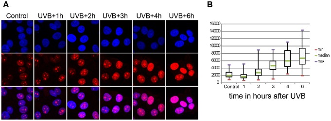
(A) Incorporation of 5-fluorouridine (5-FU) in nascent transcripts was assessed in untreated or UV irradiated cells by indirect immunofluorescence using an anti 5-FU antibody. Cell nuclei were counterstained using DAPI and the overlay between the DAPI and 5-FU signal is shown. (B) 5-FU signal intensity in the nucleus (excluding the nucleolar signal) was measured in n randomly chosen cell nuclei using the ImageJ software (NIH), where n is 59 in control, 54 in UVB+1 h, 84 in UVB+2 h, 69 in UVB+3 h, 58 in UVB+4 h, 57 in UVB+6 h. Results are presented as a boxplot where the min, max and median values are in red, purple and green respectively. P values (non-equal variance), calculated by comparing the non-irradiates sample (control) with the other time points following UVB irradiation, are the following: UVB+1 h 0,00126; UVB+2 h 0,03203; UVB+3 h 1,91441 E-13; UVB+4 h 5,72028 E-17; UVB+6 h 9,452619 E-20.