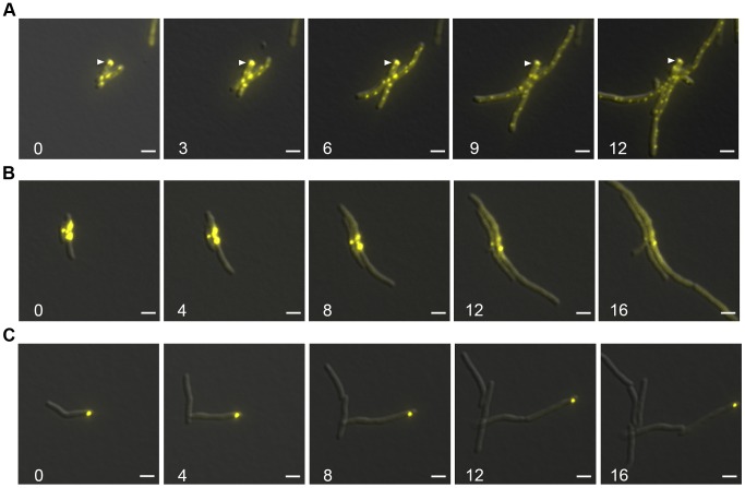
Figure 7. Protein aggregates persist during outgrowth of cells and are partitioned away from growing cells.
(A) Large static foci of DnaK-mCitrine persist during outgrowth of cells from stationary phase. Time-lapse microscopy of the DnaK-mCitrine strain (MGM6003). Time in white indicates hours after start of outgrowth. White arrow indicates large static foci that persists for over 12 hours. (B) Luciferase-mCitrine aggregates persist after growth is restored by DnaK expression. Outgrowth Tet-DnaK Luciferase-mCitrine strain (MGM6010) in the presence of ATc after DnaK depletion-induced bacteriostasis. Numbers indicate the time in hours after start of outgrowth. (C) Citrine-ELK16 aggregates persist during outgrowth. Time-lapse microscopy of aggregates during outgrowth after withdrawl of the ATc inducer of Tet-mCitrine-ELK16 expressing strain (MGM6022). For all images: Overlay of YFP and DIC images shown. White bars indicate 2 µm. Exposure times were YFP 250 ms.

