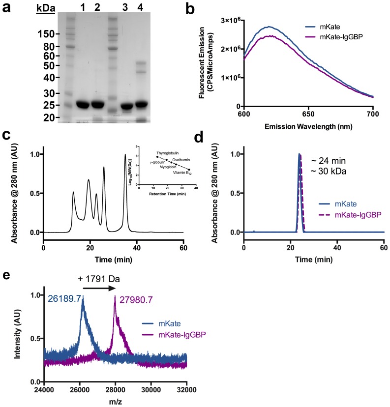
Figure 2. Characterization of mKate and mKate-IgGBP fusion.
(a) SDS-PAGE analysis of purified mKate (lanes 1, 3) and mKate-IgGBP (lanes 2, 4) under reducing (lanes 1, 2) and non-reducing conditions (lanes 3, 4). 7.5 ug of protein was loaded in each lane. (b) Fluorescence emission spectra comparing equal molar concentrations (500 nM in D-PBS) of mKate and mKate-IgGBP. (c) Size exclusion chromatography standards and associated standard curve. The standards include thyroglobulin 670 kDa, gamma-globulin 158 kDa, ovalbumin 44 kDa, myoglobin 17 kDa, and vitamin B12 1.35 kDa. (d) Size exclusion chromatogram of mKate and mKate-IgGBP with retention time and calculated molecular weight. (e) MALDI-TOF analysis of mKate and mKate-IgGBP intact mass indicating a shift in molecular weight corresponding to the expected mass of the added flexible linker and IgGBP sequence.

